Is chronic fatigue syndrome all in your brain?

Chronic fatigue syndrome (CFS) –– or myalgic encephalomyelitis/chronic fatigue syndrome (ME/CFS), to be specific –– is an illness defined by a group of symptoms. Yet medical science always seeks objective measures that go beyond the symptoms people report.
A new study from the National Institutes of Health (NIH) has performed more diverse and extensive biological measurements of people experiencing CFS than any previous research. Using immune testing, brain scans, and other tools, the researchers looked for abnormalities that might drive health complaints like crushing fatigue and brain fog. Let’s dig into what they found and what it means.
What was already known about chronic fatigue syndrome?
In people with chronic fatigue syndrome, there are underlying abnormalities in many parts of the body: The brain. The immune system. The way the body generates energy. Blood vessels. Even in the microbiome, the bacteria that live in the gut. These abnormalities have been reported in thousands of published studies over the past 40 years.
Who participated in the NIH study?
Published in February in Nature Communications, this small NIH study compared people who developed chronic fatigue syndrome after having some kind of infection with a healthy control group.
Those with CFS had been perfectly healthy before coming down with what seemed like just a simple “flu”: sore throat, coughing, aching muscles, and poor energy. However, unlike their experiences with past flulike illnesses, they did not recover. For years, they were left with debilitating fatigue, difficulty thinking, a flare-up of symptoms after exerting themselves physically or mentally, and other symptoms. Some were so debilitated that they were bedridden or homebound.
All the participants spent a week at the NIH, located outside of Washington, DC. Each day they received different tests. The extensive testing is the great strength of this latest study.
What are three important findings from the study?
The study had three key findings, including one important new discovery.
First, as was true in many previous studies, the NIH team found evidence of chronic activation of the immune system. It seemed as if the immune system was engaged in a long war against a foreign microbe — a war it could not completely win and therefore had to keep fighting.
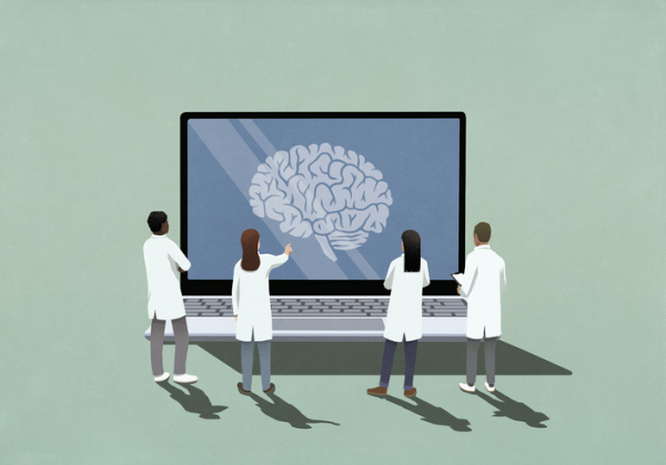
Second, the study found that a part of the brain known to be important in perceiving fatigue and encouraging effort — the right temporal-parietal area — was not functioning normally. Normally, when healthy people are asked to exert themselves physically or mentally, that area of the brain lights up during an MRI. However, in the people with CFS it lit up only dimly when they were asked to exert themselves.
While earlier research had identified many other brain abnormalities, this one was new. And this particular change makes it more difficult for people with CFS to exert themselves physically or mentally, the team concluded. It makes any effort like trying to swim against a current.
Third, in the spinal fluid, levels of various brain chemicals called neurotransmitters and markers of inflammation differed in people with CFS compared with the healthy comparison group. The spinal fluid surrounds the brain and reflects the chemistry of the brain.
What else did study show?
There are some other interesting findings in this study. The team found significant differences in many biological measurements between men and women with chronic fatigue syndrome. This surely will lead to larger studies to verify these gender-based differences, and to determine what causes them.
There was no difference between people with CFS and the healthy comparison group in the frequency of psychiatric disorders — currently, or in the past. That is, the symptoms of the illness could not be attributed to psychological causes.
Is chronic fatigue syndrome all in the brain?
The NIH team concluded that chronic fatigue syndrome is primarily a disorder of the brain, perhaps brought on by chronic immune activation and changes in the gut microbiome. This is consistent with the results of many previous studies.
The growing recognition of abnormalities involving the brain, chronic activation (and exhaustion) of the immune system, and of alterations in the gut microbiome are transforming our conception of CFS –– at least when caused by a virus. And this could help inform potential treatments.
For example, the NIH team found that some immune system cells are exhausted by their chronic state of activation. Exhausted cells don’t do as good a job at eliminating infections. The NIH team suggests that a class of drugs called immune checkpoint inhibitors may help strengthen the exhausted cells.
What are the limitations of the study?
The number of people who were studied was small: 17 people with ME/CFS and 21 healthy people of the same age and sex, who served as a comparison group. Unfortunately, the study had to be stopped before it had enrolled more people, due to the COVID-19 pandemic.
That means that the study did not have a great deal of statistical power and could have failed to detect some abnormalities. That is the weakness of the study.
The bottom line
This latest study from the NIH joins thousands of previously published scientific studies over the past 40 years. Like previous research, it also finds that people with ME/CFS have measurable abnormalities of the brain, the immune system, energy metabolism, the blood vessels, and bacteria that live in the gut.
What causes all of these different abnormalities? Do they reinforce each other, producing spiraling cycles that lead to chronic illness? How do they lead to the debilitating symptoms of the illness? We don’t yet know. What we do know is that people are suffering and that this illness is afflicting millions of Americans. The only sure way to a cure is studies like this one that identify what is going wrong in the body. Targeting those changes can point the way to effective treatments.
About the Author

Anthony L. Komaroff, MD, Editor in Chief, Harvard Health Letter
Dr. Anthony L. Komaroff is the Steven P. Simcox/Patrick A. Clifford/James H. Higby Professor of Medicine at Harvard Medical School, senior physician at Brigham and Women’s Hospital in Boston, and editor in chief of the Harvard … See Full Bio View all posts by Anthony L. Komaroff, MD

Ready to give up the lead vest?

At a dental appointment last month, I spotted a lead vest hanging unassumingly on the wall of the exam room as soon as I walked in. “Still there, but now obsolete,” I thought.
I’d just learned about new guidelines from the American Dental Association (ADA) saying lead vests and thyroid collars that cover the neck are no longer needed during dental x-rays. But they’d been a fixture of my dental experiences — including many cavities, four root canals, a tooth extraction, and two crowns — for my entire life. What changed, and could I feel safe without the vest?
Why were lead vests used in past years?
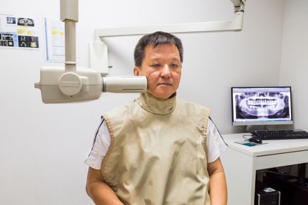
Lead vests and thyroid collars have been worn by countless Americans during dental x-rays over the years. They’ve been in use for far longer than my lifetime — about 100 years. The heavy apron-like shields are placed over sensitive areas, including the chest and neck, before the x-rays are taken.
“I haven’t worn a lead apron in the last 10 or 15 years — unless a dentist insists I put it on — because I know it isn’t needed,” says Dr. Bernard Friedland, an associate professor of oral medicine, infection, and immunity at Harvard School of Dental Medicine.
What has changed about dental x-rays?
When lead vests and thyroid collars were first recommended, x-ray technology was much less precise. But the technology has evolved significantly over the last few decades in ways that dramatically improve patient safety:
- Digital x-rays enable far smaller radiation doses, reducing radiation exposure and the risks associated with higher doses, such as cancer. “The doses used in dental radiology are negligibly small now. If you go to the dentist today for a full series of mouth x-rays that are taken with a digital sensor, the total exposure time is just over five seconds,” explains Dr. Friedland, an expert in oral radiology. “A hundred or so years ago, that exposure time would have been many minutes.”
- The small size of today’s x-ray beam significantly reduces radiation “scatter” and restricts the beam size to only the area needing to be imaged. This protects patients from radiation exposure to other parts of the body.
A less-recognized strike against using lead vests and thyroid collars is their ability to get in the way. They may block the primary x-ray beam, preventing dentists from capturing needed images. This quirk can lead to repeat imaging and unnecessary exposure to additional radiation. This is more likely to occur with panoramic x-rays.
The gear may also spread germs, Dr. Friedland notes. Although disinfected, it’s not sterilized between uses. “There’s a risk of spreading bacteria and viruses,” he says. “To me, that’s also an issue and another reason I don’t want to use one on myself.”
Who no longer needs the shields?
No one does — even children, who presumably have a long life of dental x-rays in front of them. The new recommendations apply to all patients regardless of age, health status, or pregnancy, the ADA says.
The recommendation to discontinue lead vests has been a long time in the making. In fact, the ADA isn’t the first professional organization to propose it. The American Association of Physicists in Medicine did so in 2019, followed by the American College of Radiology in 2021 and the American Academy of Oral and Maxillofacial Radiology in 2023.
Are some people confused or concerned about the no-lead-vest policy?
Yes. The new guidelines are bound to draw confusion and fear, Dr. Friedland says. Some people may even insist on continuing to wear a lead vest during x-rays.
“A big problem is that people’s perception of risk is very skewed,” he says. “Some people, you’ll never convince.”
People are likely to feel more comfortable if the practice is uniformly adopted by dentists. However, the ability to implement this change may hinge partly on public response. And it could take a while to fully adopt.
“I think the public is going to have more say on this than dentists,” Dr. Friedland says. “It might take a generation to make this change, maybe longer.”
Still concerned about the new recommendations?
If you have lingering concerns about the new recommendations, talk to your dentist.
And ask if dental x-rays are necessary to proceed with your diagnosis or treatment plan. Sometimes it’s possible to take fewer x-rays — such as bitewing x-rays of the upper and lower back teeth only — or to use certain types of imaging less frequently. Even with far safer x-ray conditions, dentists should be able to justify that the information from images is integral to diagnose problems or improve care, Dr. Friedland says.
It’s worth noting that the dose of radiation, while far lower than in the past, varies with the type of imaging and which parts of the jaw are being imaged. For example, the digital dental x-rays mentioned above involve less radiation than conventional dental x-rays. Either panoramic dental x-rays, or 3-D dental x-rays taken with a CBCT system that rotates around the head, typically involve more radiation than conventional dental x-rays.
Whenever possible, dentists should use images taken during previous dental exams, according to the ADA. “If I don’t need an x-ray, I don’t get one,” says Dr. Friedland. “I’m not cavalier about it. I also use technical parameters that keep the x-ray dose as low as reasonably possible.”
About the Author

Maureen Salamon, Executive Editor, Harvard Women's Health Watch
Maureen Salamon is executive editor of Harvard Women’s Health Watch. She began her career as a newspaper reporter and later covered health and medicine for a wide variety of websites, magazines, and hospitals. Her work has … See Full Bio View all posts by Maureen Salamon
About the Reviewer

Howard E. LeWine, MD, Chief Medical Editor, Harvard Health Publishing
Dr. Howard LeWine is a practicing internist at Brigham and Women’s Hospital in Boston, Chief Medical Editor at Harvard Health Publishing, and editor in chief of Harvard Men’s Health Watch. See Full Bio View all posts by Howard E. LeWine, MD

Does sleeping with an eye mask improve learning and alertness?

All of us have an internal clock that regulates our circadian rhythms, including when we sleep and when we are awake. And light is the single most important factor that helps establish when we should feel wakeful (generally during the day) and when we should feel sleepy (typically at night).
So, let me ask you a personal question: just how dark is your bedroom? To find out why that matters — and whether sleeping in an eye mask is worthwhile — read on.
How is light related to sleep?
Our circadian system evolved well before the advent of artificial light. As anyone who has been to Times Square can confirm, just a few watts of power can trick the brain into believing that it is daytime at any time of night. So, what’s keeping your bedroom alight?
- A tablet used in bed at night to watch a movie is more than 100 times brighter than being outside when there is a full moon.
- Working on or watching a computer screen at night is about 10 times brighter than standing in a well-lit parking lot.
Light exposure at night affects the natural processes that help prepare the body for sleep. Specifically, your pineal gland produces melatonin in response to darkness. This hormone is integral for the circadian regulation of sleep.
What happens when we are exposed to light at night?
Being exposed to light at night suppresses melatonin production, changing our sleep patterns. Compared to sleeping without a night light, adults who slept next to a night light had shallower sleep and more frequent arousals. Even outdoor artificial light at night, such as street lamps, has been linked with getting less sleep.
But the impact of light at night is not limited to just sleep. It’s also associated with increased risk of developing depressive symptoms, obesity, diabetes, and high blood pressure. Light exposure misaligned with our circadian rhythms — that is, dark during the day and light at night — is one reason scientists believe that shift work puts people at higher risk for serious health problems.
Could sleeping with an eye mask help?
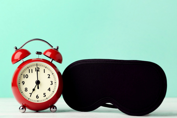
Researchers from Cardiff University in the United Kingdom conducted a series of experiments to see if wearing an eye mask while sleeping at night could improve certain measures of learning and alertness.
Roughly 90 healthy young adults, 18 to 35 years of age, alternated between sleeping while wearing an eye mask or being exposed to light at night. They recorded their sleep patterns in a sleep diary.
In the first part of the study, participants wore an intact eye mask for a week. Then during the next week, they wore an eye mask with a hole exposing each eye so that the mask didn't block the light.
After sleeping with no light exposure (wearing the intact eye mask) and with minimal light exposure (the eye mask with the holes), participants completed three cognitive tasks on days six and seven of each week:
- First was a paired-associate learning task. This helps show how effectively a person can learn new associations. Here the task was learning related word pairs. Participants performed better after wearing an intact eye mask during sleep in the days leading up to the test than after being exposed to light at night.
- Second, the researchers administered a psychomotor vigilance test, which assesses alertness. Blocking light at night also improved reaction times on this task.
- Finally, a motor skill learning test was given, which involved tapping a five-digit sequence in the correct order. For this task, there was no difference in performance whether participants had worn an intact eye mask or been exposed to light at night.
What else did the researchers learn?
No research study is ever perfect, so it is important to take the conclusions above with a grain of salt.
According to sleep diary data, there was no difference in the amount of sleep, nor in their perceptions of sleep quality, regardless of whether people wore an eye mask or not.
Further, in a second experiment with about 30 participants, the researchers tracked sleep objectively using a monitoring device called the Dreem headband. They found no changes to the structure of sleep — for example, how much time participants spent in REM sleep — when wearing an eye mask.
Should I rush out to buy an eye mask before an important meeting or exam?
If you decide to try using an eye mask, you probably don’t need to pay extra for overnight shipping. Instead, follow a chronobiologist’s rule of thumb: “bright days, dark nights.”
- During the daytime, get as much natural daylight as you possibly can: go out to pick up your morning bagel from a local bakery, take a short walk during your afternoon lull at work.
- In the evening, reduce your exposure to electronic devices such as your cell phone, and use the night-dimming modes on these devices. Make sure that you turn off all unnecessary lights. Finally, try to make your bedroom as dark as possible when you go to bed. This could mean turning the alarm clock next to your bed away from you or covering up the light on a humidifier.
Of course, you might decide a well-fitted, comfortable eye mask is a useful addition to your light hygiene toolkit. Most cost $10 to $20, so you may find yourself snoozing better and improving cognitive performance for the price of a few cups of coffee.
About the Author

Eric Zhou, PhD, Contributor
Eric Zhou, PhD, is an assistant professor at Harvard Medical School. His research focuses on how we can better understand and treat sleep disorders in both pediatric and adult populations, including those with chronic illnesses. Dr. … See Full Bio View all posts by Eric Zhou, PhD